
Knee
- Anatomy
- Conditions
- Procedures
Knee Anatomy
The knee is a complex joint made up of different structures including bones, tendons, ligaments and muscles. They all work together to maintain normal function and provide stability to the knee during movement.
Having a well-functioning healthy knee is essential for our mobility and ability to participate in various activities. Understanding the anatomy of the knee enhances your ability to discuss and choose the right treatment procedure for knee problems with Dr Maor.
Bones of the Knee
The knee is a hinge joint made up of two bones, the thighbone (femur) and the shinbone (tibia). There are two round knobs at the end of the femur called femoral condyles which articulate with the flat surface of the tibia called the tibial plateau. The tibia plateau on the inside of the leg is called the medial tibial plateau, and on the outside of the leg it is called the lateral tibial plateau.
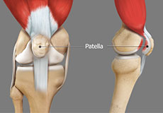
The two femoral condyles form a groove on the front (anterior) side of the knee called the patellofemoral groove. A small bone called the patella sits in this groove and forms the kneecap. It acts as a shield and protects the knee joint from direct trauma.
A fourth bone called the fibula is the other bone of the lower leg. This forms a small joint with the tibia. This joint has very little movement and is not considered a part of the main joint of the knee.
Articular Cartilage and Menisci of the Knee
Movement of the bones causes friction between the articulating surfaces. To reduce this friction, all articulating surfaces involved in movement are covered with a white, shiny, slippery layer called articular cartilage. The articulating surface of the femoral condyles, tibial plateaus and the back of the patella are covered with this cartilage. The cartilage provides a smooth surface that facilitates easy movement.
To further reduce friction between the articulating surfaces of the bones, the knee joint is lined by a synovial membrane which produces a thick clear fluid called synovial fluid. This fluid lubricates and nourishes the cartilage and bones inside the joint capsule.
Within the knee joint, between the femur and tibia, there are two C shaped cartilaginous structures called menisci. Menisci function to provide stability to the knee by spreading the weight of the upper body across the whole surface of the tibial plateau. The menisci help in load- bearing by preventing the weight from concentrating onto a small area, which could damage the articular cartilage. The menisci also act as a cushion between the femur and tibia by absorbing the shock produced by activities such as walking, running and jumping.
Ligaments of the Knee
Ligaments are tough bands of tissue that connect one bone to another bone. The ligaments of the knee function to stabilize the knee joint. There are two important groups of ligaments that hold the bones of the knee joint together, collateral ligaments and the cruciate ligament.
Collateral ligaments are present on either side of the knee. They function to prevent the knee from moving too far during side to side motion. The collateral ligament on the inside is called the medial collateral ligament (MCL) and the collateral ligament on the outside is called the lateral collateral ligament (LCL).
Cruciate ligaments, present inside the knee joint, control the back-and-forth motion of the knee. The cruciate ligament in the front of the knee is called anterior cruciate ligament or ACL and the cruciate ligament in the back of the knee is called posterior cruciate ligament or PCL.
Muscles of the Knee
Muscles: There are two major muscles, the quadriceps and the hamstrings, which enable movement of the knee joint. The quadriceps muscles are in the front of the thigh. When the quadriceps muscles contract, the knee straightens. The hamstrings are in the back of the thigh. When the hamstring muscles contract, the knee bends.
Tendons of the Knee
Tendons are structures that attach muscles to the bone. The quadriceps muscles of the knee meet just above the patella and attach to it through a tendon called the quadriceps tendon. The patella further attaches to the tibia through a tendon called the patella tendon. The quadriceps muscle, quadriceps tendon and patellar tendon all work together to straighten the knee. Similarly, the hamstring muscles at the back of the leg are attached to the knee joint with the hamstring tendon.
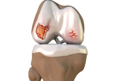
Osteoarthritis
Arthritis is a general term covering numerous conditions where the joint surface or cartilage wears out. The joint surface is covered by a smooth articular surface that allows pain-free movement in the joint. This surface can wear out for several reasons; often the definite cause is not known. Arthritis often affects the knee joint.
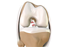
ACL Tears & Sprains
An ACL injury is a sports-related injury that occurs when the knee is forcefully twisted or hyperextended. An ACL tear usually occurs with an abrupt directional change with the foot fixed on the ground or when the deceleration force crosses the knee.
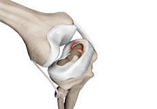
Meniscal Tears
Meniscal tears often occur during sports. These tears are usually caused by twisting motion or over flexing of the knee joint. Athletes who play sports such as football, tennis and basketball are at a higher risk of developing meniscal tears. They often occur along with injuries to the anterior cruciate ligament, a ligament that crosses from the femur (thighbone) to the tibia (shinbone).
Patellofemoral Instability
The knee can be divided into three compartments: patellofemoral, medial and lateral compartment. The patellofemoral compartment is the compartment in the front of the knee between the kneecap and thighbone.
Runner’s Knee
Patellofemoral pain syndrome also called runner’s knee refers to pain under and around your kneecap. Patellofemoral pain is seen in number of medical conditions such as anterior knee pain syndrome, patellofemoral malalignment, and chondromalacia patella that cause pain around the front of the knee.
Osteochondral Defects (OCD)
Osteochondral defects of the knee are segments of the knee cartilage and bone which detaches from the underlying bone. This can cause pain, swelling, clicking, catching and locking of the knee. They typically affect young people, and more commonly men than women.
Fractures & Trauma
A bone fracture is a medical condition in which a bone is cracked or broken. It is a break in the continuity of the bone. While many fractures are the result of high force impact or stress, bone fracture can also occur because of certain medical conditions that weaken the bones, such as osteoporosis.
Other Knee Conditions
The other knee conditions include knee pain, patellar tendinitis, chondromalacia patella, lateral patellar compression, patellofemoral dislocation, Osgood schlatter disease, Jumper's knee, bursitis, Baker's cyst and iliotibial band syndrome.
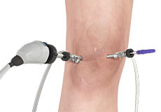
Knee Arthroscopy
Knee Arthroscopy is a common surgical procedure performed using an arthroscope, a viewing instrument, to diagnose or treat a knee problem. It is a relatively safe procedure and most of the patients are discharged from the hospital on the same day of surgery.
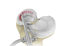
Meniscal Surgery
A meniscus tear is the most common knee injury in athletes, especially those involved in contact sports. A sudden bend or twist in your knee can cause the meniscus to tear. This is a traumatic meniscal tear. Elderly people are more prone to degenerative meniscal tears as the cartilage wears out and weakens with age.
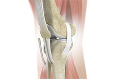
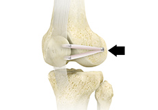
ACL Reconstruction
The anterior cruciate ligament is one of the major stabilizing ligaments in the knee. It is a strong rope- like structure located in the center of the knee running from the femur to the tibia. When this ligament tears unfortunately, it does not heal and often leads to the feeling of instability in the knee.
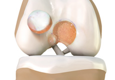
Cartilage Reconstruction
The common techniques employed for cartilage restoration include cartilage replacement and cartilage repair & transplantation.
Cartilage replacement is a surgical procedure performed to replace the worn-out cartilage with new cartilage. It is usually performed to treat patients with small areas of cartilage damage usually caused by sports or traumatic injuries. It is not indicated for those patients who have advanced arthritis of the knee.
Patellofemoral Stabilisation
The knee can be divided into three compartments: patellofemoral, medial and lateral compartment. The patellofemoral compartment is the compartment in the front of the knee between the knee cap and thigh bone. The medial compartment is the area on the inside portion of the knee, and the lateral compartment is the area on the outside portion of the knee joint.
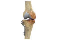
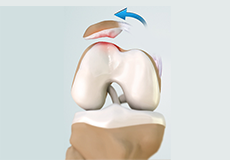
High Tibial Osteotomy
High tibial osteotomy is a surgical procedure performed to relieve pressure on the damaged site of an arthritic knee joint. It is usually performed in arthritic conditions affecting only one side of your knee and the aim is to take pressure off the damaged area and shift it to the other side of your knee with healthy cartilage.
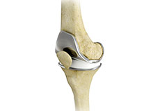
Partial Knee Replacement
Arthritis is inflammation of a joint causing pain, swelling (inflammation), and stiffness.
Osteoarthritis is the most common form of knee arthritis in which the joint cartilage gradually wears away. It most often affects older people. In a normal joint, articular cartilage allows for smooth movement within the joint, where as in an arthritic knee the cartilage itself becomes thinner or completely absent.
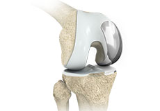
Total Knee Replacement
Total knee replacement, also called total knee arthroplasty, is a surgical procedure in which the worn out or damaged surfaces of the knee joint are removed and replaced with artificial parts.
Total knee replacement surgery is commonly indicated for severe osteoarthritis of the knee. Osteoarthritis is the most common form of knee arthritis in which the joint cartilage gradually wears away. It often affects older people.