Paediatrics
- Paediatric Knee Conditions
- Paediatric Foot Conditions
- Paediatric Fractures
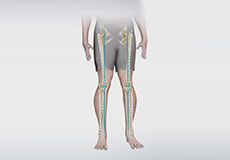
Knock Knees and Bow Legs
Angular deformities of the knee are common during childhood and usually are variations in the normal growth pattern. Angular deformity of the knee is a part of normal growth and development during early childhood.
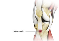
Osgood Schlatter Disease
Osgood-Schlatter disease refers to a condition of an overuse injury that occurs in the knee region of growing children and adolescents. This is caused by inflammation of the tendon located below the kneecap (patellar tendon). Children and adolescents who participate in sports such as soccer, gymnastics, basketball and distance running are at higher risk of this disease.
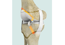
Paediatric ACL Reconstruction
Tibial spine avulsion is tearing away of the tibial eminence (an extension on the bone for attachment of muscles) which involves the anterior cruciate ligament (ACL) insertion site.
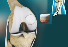
Osteochondritis Dissecans (OCD)
Osteochondral defects of the knee are segments of the knee cartilage and bone which detaches from the underlying bone. This can cause pain, swelling, clicking, catching and locking of the knee. They typically affect young people, and more commonly men than women.
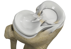
Discoid Meniscus
The medial meniscus is on the inside of the knee, it is attached to the ligaments on the inside of the knee (MCL) and the joint capsule. As it is more firmly attached to the deep structures of the knee, it is more commonly injured than the lateral meniscus. It takes approximately 50% of the weight across the medial (inside) compartment of the knee.
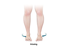
Intoeing
In toeing, also called “pigeon-toed”, is an abnormal condition characterized by inward facing of the toe or foot instead of being straight. Parents may observe intoeing in their children at an early age when they start walking. But usually intoeing corrects itself without any specific treatment as the child grows up to around 8 years of age.
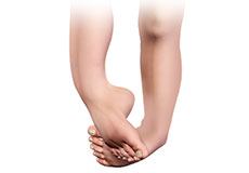
Clubfoot
Congenital clubfoot is a common pediatric foot deformity. The feet twist inward and downward at the ankles in such a way that the ankle or side of the foot meets the ground while walking instead of the sole of the foot. It is twice as common in males as females. The leg and foot may be smaller and calves less developed than normal.
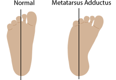
Metatarsus Adductus
Metatarsus adductus is a paediatric foot deformity. This is a condition where the foot of the child may bend inward from the middle to the toes. This condition is similar to clubfoot deformity.
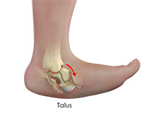
Vertical Talus
Congenital vertical talus (CVT) is a rare condition in which the talus (heel bone) and navicular bones (ankle bone) of the child’s feet are abnormally positioned. This leads to a rigid flat foot with a rocker-bottom appearance. The hindfoot points downward to the floor while the forefoot points upwards. It occurs most frequently with other neuromuscular disorders such as spina bifida and arthrogryposis (multiple joint contractures present at birth).
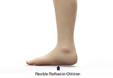
Paediatric Flexible Flatfoot
Flatfoot, also known as “fallen arches” or Pes planus, is a deformity in children’s feet in which the arch that runs lengthwise along the sole of the foot has collapsed to the ground or not formed at all. Flatfoot is normal in the first few years of life as the arch of the foot usually develops between the age of 3 and 5 years.
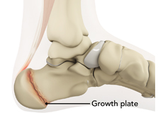
Sever's Disease
Sever's disease is a painful inflammation of the growth plate in the heel. Growth plates are areas at the end of children’s bones that undergo changes so bone growth can occur. During this time, the muscles and tendons may not grow as fast as the bone causing tightness and pressure at the back of the heel.
Fractures/Broken bones in children are very common. Children who are still growing have different anatomy, they are not ‘mini adults’ and therefore their fractures need special consideration and treatment.
ANATOMY
Children have growth plates in their bones, where new growth is formed. If these are injured it can have long lasting consequences on their growth and soft tissue development if not treated appropriately. Children’s bones also are softer and more elastic than adults, this makes it much easier to bend rather than break/fracture. Similarly, children’s bones are covered in a very thick layer of supporting tissue called periosteum, which assists in fracture healing, and means that they recover much faster than adults’. Children also have great potential to ‘remodel’ or ‘grow out’ deformity from fractures, which adults cannot do.
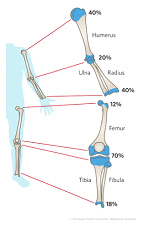
Growth plates of the body and how much they contribute to limb length
If your child has a growth plate fracture they may show symptoms such as:
- Difficulty in moving the limb in the affected area
- Pain and tenderness of the affected bone
- Difficulty in carrying weight or putting pressure on the affected limb
- Swelling and warmth near the joint
Dr Maor may diagnose growth plate fractures with the help of imaging tests such as X-ray, CT scan and MRI.
Treatment for growth plate fractures depends on the severity of the fracture.
NON SURGICAL TREATMENT
The majority of children’s fractures can be treated without open surgery. Non-surgical treatment commonly involves:
- Casts
- Braces/Splints
- Boots
Due to the remodelling potential of children, even if the bones are not perfectly aligned, over time as the child grows they will straighten out, this is known as “remodelling”. This process can take years to be complete. The key considerations for Dr Maor to decide whether or not a fracture will remodel sufficiently are:
- Age of the child
- Amount of displacement
- Which bone is affected
- Which direction the bones have shifted
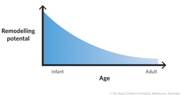
The rate of remodelling in a child is inversely related to age. In younger children, remodelling is rapid and often complete. It is slower in older children and much slower in adolescents
Fracture and bone remodelling potential with age

Right Femur fracture showing remodelling (straightening) over time)
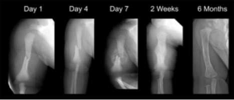
Humerus (arm bone) showing remodelling over time
SURGICAL TREATMENT
Surgical treatment is sometimes required for children’s fractures to assist in realigning bones and joints to allow effective healing to occur. This may involve:
- Pushing the bones back into place under anaesthetic, without opening the skin/without incisions
- Mini open (keyhole) manipulation and casts
- Closed/Open reduction with fixation

